
Services Overview
While most first-time visits to Triton Eye Care will be for a “routine eye exam,” our service is beyond routine. All Patients presenting without an emergent or acute eye problem undergo a Comprehensive Eye Health Evaluation. Dr. Gary Morgan has years of experience in treating typical vision disorders such as nearsightedness, farsightedness, astigmatism, and presbyopia. But more importantly, your overall visual and eye health are assessed. Our advanced diagnostic equipment tests not only the quantity of your vison (your visual acuity) but also the quality of your vision, known as Contrast Sensitivity. The health of your entire visual system is evaluated, looking for medical eye conditions such as Eye allergies, blepharitis, dry eyes, cataracts, vitreous degeneration (floaters), glaucoma, macular pucker, or Age-related macular degeneration (AMD). And if you have a systemic health condition such as diabetes or hypertension or are on a high-risk medication such as hydroxychloroquine (Plaquenil™), Dr. Morgan will specifically examine for their ocular side effects and send a detailed report to your primary care physician. Our practice also offers testing to evaluate your risk of eye disease.
Of course, life happens and your first visit to Triton Eye Care may be for an emergency such as a foreign body, an eye infection, or a red, painful eye. Rest assured that eye emergencies are always seen same day. If you are experiencing an eye emergency outside of normal business hours, please call our office and leave a message, we will return your call as soon as possible!
Automated Vision Testing
Which is better?
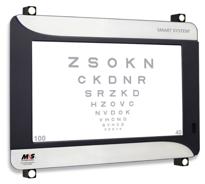
Refraction is the part of the eye exam when vision is tested, and a glasses prescription is determined. For many patients, this can be frustrating or even nerve-wracking! At Triton eye Care, we use the latest automated vision testing equipment. This eases the testing procedure and minimizes the decisions you need to make during the exam. Many of the traditional choices, “which is better, one or two,” are replaced with simultaneous images being displayed to make your choice. In other words, you don’t have to remember what “choice one” looks like when comparing it to “choice two,” they are both seen at the same time!
Bring Your Glasses
Now, the thing about automated vision testing is sometimes it can be too accurate. While one may test the same way each time, the resultant prescription can be uncomfortable due to familiarity with one’s current prescription. This can especially be true with astigmatism correction. Therefore, please be sure to bring all of your current pairs of glasses to your exam, even if you don’t like the vision through one or all of them. The prescription in your glasses will be read by our equipment so a comparison can be made with your test results, and a determination can be made by Dr. Morgan to give you your best possible visual outcome. Your finest, most comfortable vision is our goal for your new prescription. Of course, if any underlying conditions that may limit vision are found during your exam, they will be thoroughly explained.
Contrast Sensitivity and Glare Testing
Dr. Gary Morgan goes beyond normal testing. Did you know that 20/20 vision is not the same for all people? Contrast Sensitivity is a measure of one’s ability to see a target against its background. This can also be referred to as ‘faintness appreciation.’ Imagine driving directly into the sun at dusk when a child steps into the road. Now, the child is much larger than a 20/20 letter on an eye chart, but individuals will vary greatly in their ability to see the child (the target) against the glaring sun (the background). One’s Contrast Sensitivity in this case could be the difference in avoidance versus tragedy.
Related to Contrast Sensitivity is one’s ability to tolerate Glare. Glare occurs when an individual is exposed to a light source, either direct or indirect, that is in excess of their adaptive state. Such light exposure can cause discomfort, disability, or both. Discomfort Glare diminishes visual performance, we can still see, just not as well. The discomfort is made worse by short wavelength / high energy light, also known as blue light. Sunlight is predominantly blue, which is why the sky is blue. So, sunlight exposure, especially when the sun is low on the horizon as at dusk or dawn will increase our discomfort. Newer high-intensity automobile headlights that appear blue can also cause Discomfort Glare.
Disability Glare occurs from reduction of contrast; one can no longer tell a visual target from its background. It can occur directly as when driving into the sun or oncoming headlights. Disability Glare can also occur indirectly, as in when our eyes are exposed to a sudden bright light or flash. This bleaches the photopigment in our retina, causing photostress, and wiping out our contrast.
 All patients receiving a vision exam at Triton Eye Care will have their Contrast Sensitivity and Glare tested. For those that don’t test up to par, specific recommendations will be made to improve their Contrast Sensitivity and Glare tolerance. For those interested in complying with recommendations, periodic follow-up testing will be performed to track improvements.
Topcon Maestro 2
Retinal Photo
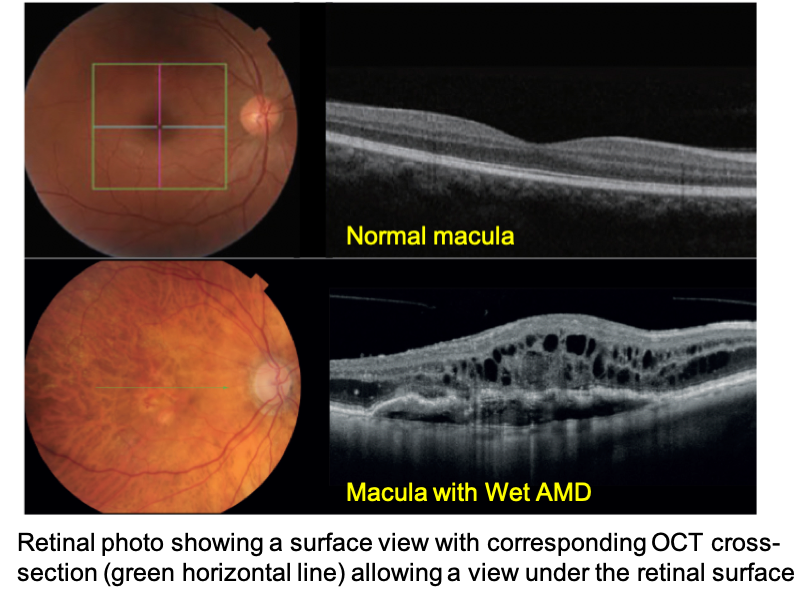 Determining with accuracy the health of the retina and optic nerve are critical in eye care. Triton Eye Care employs the Topcon Maestro 2 for detailed imaging of the inside of the eye. Two types of imaging are obtained simultaneously with the Maestro. First, a photo of the retina is taken, which allows for a surface view of the back of the eye. While Dr. Morgan also takes a live look at the back of your eye during an exam, the views can only be momentary, or the light used would become uncomfortable. With the retinal photo, a quick flash of light captures an image of the back of the eye that can be more easily studied. The photo can also be used to educate you on your eye health. The photo is stored in your medical record for reference, allowing for easy future comparison.
OCT
The second type of imaging captured is called Optical Coherence Tomography, or OCT. An OCT image allows what lies beneath the surface to be seen as well. Why is this important? Well, imagine this scenario…
You are navigating a boat down a jungle river. You come around a bend and see a tree sticking out of the water. You don’t want to hit the tree, so you steer the boat in an attempt to get around it. What you can’t see is that the tree actually fell from shore, and you steered the boat right into its thick trunk, which is submerged below the water. Hitting the trunk, the boat capsizes, and you are thrown from the boat into the water. Being in the jungle, there are all kinds of hungry, dangerous creatures such as piranhas and crocodiles in the water. If only you could have seen below the surface of the water! Certainly, you would have steered the boat around the opposite side of the tree…
Hence, the importance of OCT technology during an eye exam. Sometimes, things may appear normal on the surface of the retina and optic nerve when in fact, they are not. Early macular degeneration, early macular edema from diabetes, early thinning of optic nerve tissue that can lead to glaucoma, and other insidious conditions can all be uncovered in a timely manner. Early detection of eye disease allows for prevention and treatment strategies to be promptly undertaken to preserve vision.
OCT is a non-invasive, non-contact procedure that doesn't require preparation from the patient. There is no exposure to radiation since unlike images obtained by an x-ray, CT scan, or MRI, the machine simply uses light to obtain the images. The patient sits in front of the OCT instrument, 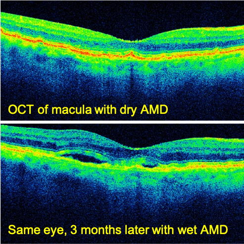 a couple of bright flashes like a normal camera flash go off, and the images are available for viewing within a minute.
OCT and AMD
For patients with Age-related macular degeneration (AMD) , OCT is an invaluable tool to monitor for the development of wet AMD. While all patients start out with the dry form of AMD, some will progress to the wet form. Wet AMD occurs when new blood vessels grow under the retina, which don’t become visible on the retinal surface until they have leaked blood and affected vision. Periodic monitoring of dry AMD with OCT allows for early detection of wet AMD, many times before the patient is aware of any visual change. When caught early, wet AMD can be effectively treated with medication to prevent further abnormal blood vessel growth, preserving vision.
OCT and Diabetes
According to the US National Institute of Health (NIH), 30.3 million people have diabetes, 24 percent of which are undiagnosed. Additionally, 107.2 million American adults have prediabetes. Many times, diabetes is discovered during an eye exam, and testing with a retinal photo and OCT (performed simultaneously with the Maestro 2 is invaluable in making the diagnosis. This allows for prompt referral to your physician for evaluation of treatment. For those diagnosed with diabetes, an essential part of your treatment is a yearly eye exam to monitor for the development or progression of diabetic retinopathy , of which the Maestro 2 is an integral part. After your exam, Dr. Morgan will send a detailed report to your doctor.
OCT and Glaucoma
Another common use of the OCT is for monitoring and managing glaucoma. Our office usually takes initial images of the optic nerve, and the OCT analyzes and compares these images to a vast database of age-matched healthy control patients. The OCT will usually be repeated every year to follow any changes or progression over time.
The advent of OCT has revolutionized the way we evaluate the retina because we can now detect subtle findings not otherwise easily seen during clinical exams. This makes the OCT one of the most valuable tests we can do in our office.