
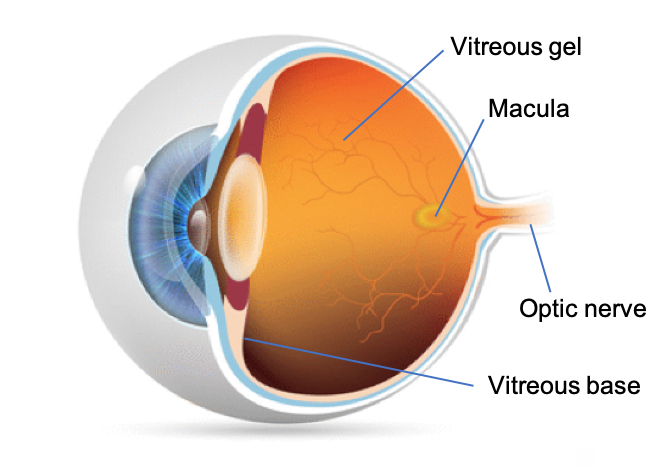
 The vitreous is a gel-like substance that fills the posterior cavity of the eye. The outer surface of the gel contains millions of tiny fibers that tightly adhere to the vitreous base anteriorly, and with a less tight adherence to the retina and optic nerve posteriorly. As we age, the gel starts to shrink, the fibers break, and the gel pulls away from the retina causing floaters. When you see floaters, you are not actually ‘seeing’ them. You are seeing the shadow of the floaters. Therefore, you will notice floaters more in bright sunshine than in a dimly lit room.
Posterior Vitreous Detachment (PVD)
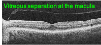 As the gel continues to shrink and pull away from the retina, one may notice a sudden increase in floaters or what appears as cobwebs in their vision. This may indicate that the vitreous has more fully separated from the retina and optic nerve as seen in the OCT photo to the right. Note the line above the retinal cross section, that is the vitreous face. In fact, an annular ring may be noticed along with floaters signifying the vitreous detachment form the optic nerve. Flashes of light that look like lightening streaks may also appear in your peripheral vision as the gel detaches and tugs on the retina.
PVD complications
Fortunately, eighty-five to ninety percent of PVDs do not result in any harm; PVD is a natural occurrence with age. However, sometimes problems do occur. First, as the fibrils pull at the vitreous base, if they don’t break, it can cause a tear in the retina. Fortunately, retinal tears can usually be treated quite easily, but time is of the essence. The sooner a retinal tear is treated, the less likely it will progress to a retinal detachment. While a retinal detachment can also be treated, the risk of vision loss is higher.
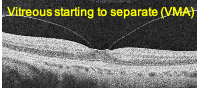 Second, the vitreous may incompletely detach from the retina posteriorly at the macula, leading to a condition known as vitreomacular adhesion (VMA). As can be seen from the OCT image on the right, a small amount of VMA may occur in early stages of PVD. The majority of the time this leads to a complete separation with no further visual consequences.
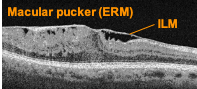 Third, sometimes as the gel separates, the vitreal fibrils don’t release easily and tug on their retinal attachment points over the macula. This leads to a wrinkling of the surface of macula, sometimes referred to as a macular pucker. A more formal name for this condition is Epiretinal membrane (ERM). The internal limiting membrane (ILM) is the outermost layer of the retina. As the fibrils pull on the ILM, it wrinkles and separates from the retina, henceforth being called an ERM. If the ERM significantly distorts vision, surgical repair is required.
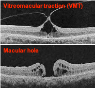 Fourth, the vitreal fibers may be more firmly attached at the macula leading to vitreomacular traction (VMT). VMT may cause visual distortion, such as a doorframe looking like it is bent. VMT is a more worrisome condition because if the vitreous and retina do not separate, the VMT can cause a macular hole. A sudden symptom of a dark spot in the center of vision may indicate a macular hole has formed. Macular holes must be surgically repaired.
Get Evaluated!